Discussions highlight the usefulness of the dataset for visualizing fine brain features and improving fMRI signals, with examples like segmentation of the LGN and blood motion artifacts. The tone emphasizes technical interest and potential scientific impact, reflecting curiosity and recognition of the dataset's relevance in neuroimaging.
AgreementModerate agreement
Most discussions recognize the significance of the dataset and methods, with some referencing prior work or ongoing research interest.
InterestHigh interest
Posts show high curiosity about the detailed imaging and potential applications, especially in studying brain structures.
EngagementHigh engagement
Participants actively discuss segmentation techniques, artifact handling, and implications for fMRI research, indicating deep engagement.
ImpactModerate impact
The conversations suggest that the dataset could advance neuroimaging understanding, though some see it as an incremental step rather than revolutionary.
Abstract in brief
The text discusses a new dataset of detailed in vivo human brain measurements using high-resolution (0.35mm) MRI at 7 Tesla, focusing on the T parameter which is important for understanding brain structure.
This dataset allows researchers to visualize fine brain features like cortical layers and blood vessels, and shows consistent depth-related T values in specific brain regions like the visual and auditory cortex.
The data is openly available online, and it can be useful for studying brain anatomy and improving the interpretation of T-weighted fMRI signals.]
Latest lab work in @NeuroImage_EiC by @ofgulban. We release high-quality mesoscopic (0.35-mm) anatomical measurements in the living human brain. Clear laminar and vascular features. May help us better understand the nature of fMRI signals. https://t.co/ndyCtLntDh https://t.co/qRcFJuJ9Rl
Nov 17, 2022 · 62 likes · 17 shares · 1,801 followersView post →
FG
Faruk Gulban
@ofgulban · X
A short tutorial on getting started with segmentation using ITKSNAP: https://t.co/6ZFWWB5vrq
Particularly, I am segmenting the LGN (lateral geniculate nucleus) of a living human brain using our publicly available 0.35 mm isotropic 7 T dataset.
Mar 21, 2023 · 17 likes · 5 shares · 638 followersView post →
TY
Thomas Yeo
@bttyeo · X
RT @cvnlab: Latest lab work in @NeuroImage_EiC by @ofgulban. We release high-quality mesoscopic (0.35-mm) anatomical measurements in the li…
Nov 18, 2022 · 17 shares · 11,820 followersView post →
AF
Alex Fornito
@AFornito · X
RT @cvnlab: Latest lab work in @NeuroImage_EiC by @ofgulban. We release high-quality mesoscopic (0.35-mm) anatomical measurements in the li…
Nov 18, 2022 · 17 shares · 7,787 followersView post →
L
layerfMRI
@layerfMRI · X
RT @cvnlab: Latest lab work in @NeuroImage_EiC by @ofgulban. We release high-quality mesoscopic (0.35-mm) anatomical measurements in the li…
Nov 17, 2022 · 17 shares · 3,789 followersView post →
RO
Rosanna Olsen
@rosanna_olsen · X
RT @cvnlab: Latest lab work in @NeuroImage_EiC by @ofgulban. We release high-quality mesoscopic (0.35-mm) anatomical measurements in the li…
Nov 18, 2022 · 17 shares · 3,240 followersView post →
This tutorial covers segmenting the lateral geniculate nucleus using high-resolution 7 Tesla MRI data, highlighting detailed in vivo human brain measurements and visualization of fine brain features. The dataset is…
This seminar discusses a new dataset of detailed in vivo human brain measurements using high-resolution MRI at 7 Tesla, focusing on the T parameter to visualize cortical layers and blood vessels, enhancing understanding…
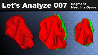
This video demonstrates the segmentation of Heschl's Gyrus, the auditory cortex region, in human brains using 0.35 mm T2* weighted MRI data acquired at 7 Tesla, highlighting detailed brain structure analysis techniques.
This video demonstrates how to crop NIfTI brain images using Python and the FSLROI program, a method essential for precise analysis of high-resolution MRI datasets, like the 7 Tesla T parameter dataset.
More on the blood motion artifact, and capturing it on 7 T MRI, see my 2022 paper: https://t.co/QDjGDFefsO
I call this artifact shortly "blood motion artifact", but in the past it was called "spatial misregistration of the vascular flow". Larson 1990 https://t.co/OngX2M7FwY
RT @ofgulban: A short tutorial on getting started with segmentation using ITKSNAP: https://t.co/6ZFWWB5vrq
Particularly, I am segmenting t…
Mar 22, 2023 · 5 shares · 678 followersView post →
K小
Kenshu Koiso / 小磯堅秀
@kenkenkenshu · X
RT @ofgulban: A short tutorial on getting started with segmentation using ITKSNAP: https://t.co/6ZFWWB5vrq
Particularly, I am segmenting t…
Mar 21, 2023 · 5 shares · 194 followersView post →
AB
Alexandra Badea
@portokalh · X
RT @ofgulban: A short tutorial on getting started with segmentation using ITKSNAP: https://t.co/6ZFWWB5vrq
Particularly, I am segmenting t…
Mar 21, 2023 · 5 shares · 958 followersView post →
BL
BAND-Lab
@BANDLabUM · X
RT @ofgulban: A short tutorial on getting started with segmentation using ITKSNAP: https://t.co/6ZFWWB5vrq
Particularly, I am segmenting t…
Mar 21, 2023 · 5 shares · 924 followersView post →
CB
Claude Bajada
@ClaudeBajada · X
RT @ofgulban: A short tutorial on getting started with segmentation using ITKSNAP: https://t.co/6ZFWWB5vrq
Particularly, I am segmenting t…
Mar 21, 2023 · 5 shares · 693 followersView post →
FG
Faruk Gulban
@ofgulban · X
A short tutorial on getting started with segmentation using ITKSNAP: https://t.co/6ZFWWB5vrq
Particularly, I am segmenting the LGN (lateral geniculate nucleus) of a living human brain using our publicly available 0.35 mm isotropic 7 T dataset.
Mar 21, 2023 · 17 likes · 5 shares · 638 followersView post →
LL
Lizzy Levarity
@LizLevarity · X
RT @cvnlab: Latest lab work in @NeuroImage_EiC by @ofgulban. We release high-quality mesoscopic (0.35-mm) anatomical measurements in the li…
RT @cvnlab: Latest lab work in @NeuroImage_EiC by @ofgulban. We release high-quality mesoscopic (0.35-mm) anatomical measurements in the li…
Mar 12, 2023 · 17 shares · 117 followersView post →
CS
Chinese Open Science @ chineseopensci.bsky.social
@ChineseOpenSci · X
RT @cvnlab: Latest lab work in @NeuroImage_EiC by @ofgulban. We release high-quality mesoscopic (0.35-mm) anatomical measurements in the li…
Nov 20, 2022 · 17 shares · 689 followersView post →
DT
Dylan Tisdall
@mdtisdall · X
RT @cvnlab: Latest lab work in @NeuroImage_EiC by @ofgulban. We release high-quality mesoscopic (0.35-mm) anatomical measurements in the li…
Nov 19, 2022 · 17 shares · 81 followersView post →
AP
Alessandra Pizzuti
@alepizz_ · X
RT @cvnlab: Latest lab work in @NeuroImage_EiC by @ofgulban. We release high-quality mesoscopic (0.35-mm) anatomical measurements in the li…
Nov 19, 2022 · 17 shares · 225 followersView post →
Y
Yasharz
@YasharZeighami · X
RT @cvnlab: Latest lab work in @NeuroImage_EiC by @ofgulban. We release high-quality mesoscopic (0.35-mm) anatomical measurements in the li…
Nov 19, 2022 · 17 shares · 626 followersView post →
UN
University of Minnesota Neuroscience
@UMNeurosci · X
RT @cvnlab: Latest lab work in @NeuroImage_EiC by @ofgulban. We release high-quality mesoscopic (0.35-mm) anatomical measurements in the li…
Nov 18, 2022 · 17 shares · 2,208 followersView post →
AK
Aneurin J Kennerley
@MagneticDr_K · X
RT @cvnlab: Latest lab work in @NeuroImage_EiC by @ofgulban. We release high-quality mesoscopic (0.35-mm) anatomical measurements in the li…
Nov 18, 2022 · 17 shares · 782 followersView post →
RO
Rosanna Olsen
@rosanna_olsen · X
RT @cvnlab: Latest lab work in @NeuroImage_EiC by @ofgulban. We release high-quality mesoscopic (0.35-mm) anatomical measurements in the li…
Nov 18, 2022 · 17 shares · 3,240 followersView post →
TY
Thomas Yeo
@bttyeo · X
RT @cvnlab: Latest lab work in @NeuroImage_EiC by @ofgulban. We release high-quality mesoscopic (0.35-mm) anatomical measurements in the li…
Nov 18, 2022 · 17 shares · 11,820 followersView post →
AF
Alex Fornito
@AFornito · X
RT @cvnlab: Latest lab work in @NeuroImage_EiC by @ofgulban. We release high-quality mesoscopic (0.35-mm) anatomical measurements in the li…
Nov 18, 2022 · 17 shares · 7,787 followersView post →
E
Elisa
@elizamb89 · X
RT @cvnlab: Latest lab work in @NeuroImage_EiC by @ofgulban. We release high-quality mesoscopic (0.35-mm) anatomical measurements in the li…
Nov 18, 2022 · 17 shares · 59 followersView post →
T
thmonk
@thmonk_pf · X
RT @cvnlab: Latest lab work in @NeuroImage_EiC by @ofgulban. We release high-quality mesoscopic (0.35-mm) anatomical measurements in the li…
Nov 17, 2022 · 17 shares · 1,633 followersView post →
LF
Lingzhong Fan
@Lingzhong_Fan · X
RT @cvnlab: Latest lab work in @NeuroImage_EiC by @ofgulban. We release high-quality mesoscopic (0.35-mm) anatomical measurements in the li…
Nov 17, 2022 · 17 shares · 596 followersView post →
FG
Faruk Gulban
@ofgulban · X
RT @cvnlab: Latest lab work in @NeuroImage_EiC by @ofgulban. We release high-quality mesoscopic (0.35-mm) anatomical measurements in the li…
Nov 17, 2022 · 17 shares · 638 followersView post →
L
layerfMRI
@layerfMRI · X
RT @cvnlab: Latest lab work in @NeuroImage_EiC by @ofgulban. We release high-quality mesoscopic (0.35-mm) anatomical measurements in the li…
Nov 17, 2022 · 17 shares · 3,789 followersView post →
C
cvnlab
@cvnlab · X
Latest lab work in @NeuroImage_EiC by @ofgulban. We release high-quality mesoscopic (0.35-mm) anatomical measurements in the living human brain. Clear laminar and vascular features. May help us better understand the nature of fMRI signals. https://t.co/ndyCtLntDh https://t.co/qRcFJuJ9Rl
Nov 17, 2022 · 62 likes · 17 shares · 1,801 followersView post →