The group discusses the innovative workflow combining light and electron microscopy, with examples like 'revealing the ultrastructure' and references to '3D ultrastructure' and 'targeting microorganisms,' showing both appreciation and interest in methodological breakthroughs. Words such as 'beautiful' and 'amazing' highlight admiration, while mentions of overcoming 'technical challenges' emphasize its potential impact.
AgreementStrong agreement
Most discussions express strong support and praise for the research's value and advancements.
InterestHigh interest
The detailed mentions of new techniques and the marine microorganism demonstrate high curiosity and engagement.
EngagementHigh engagement
Discussion includes technical insights, workflow improvements, and visualizations, indicating deep involvement.
ImpactHigh impact
The focus on methodological validation and environmental applications suggests the research could significantly influence microbiology and microscopy fields.
Abstract in brief
The study presents a method combining light and electron microscopy to isolate and examine a specific marine microorganism, Ensiculifera tyrrhenica, within its natural, complex environment.
Using this approach, researchers detailed the internal structure of the dinoflagellate, including its single complex chloroplast, flagellar apparatus, and potential chromatin activity, providing insights into its cell biology.
This workflow demonstrates an effective way to study individual microorganisms in environmental samples, overcoming technical challenges posed by sample heterogeneity and low throughput of traditional volume electron microscopy.
The final version of our manuscript on Targeted volume correlative light and electron microscopy of an environmental marine microorganism is out in JCS
@MocaerK @SchwabYannick @VolumeEM1 https://t.co/PUBKsSb7Ag
New #microscopy method can help put sharper focus on #plankton samples https://t.co/1txOyONnfn https://t.co/v6BAH5bx6I
Oct 5, 2023 · 32 likes · 13 shares · 1,022,459 followersView post →
F
FocalPlane
@focalplane_jcs · X
This #EMmonday we are featuring the beautiful vCLEM paper from @MocaerK @SchwabYannick @PaoloRonchi10, et al. They reveal the ultrastructure of a marine dinoflagellate, validating their method for targeting microorganisms from heterogenous field samples.
https://t.co/l3IAruUiBz https://t.co/tIxVwH3Beu
Aug 14, 2023 · 28 likes · 7 shares · 5,963 followersView post →
JS
J Cell Science
@J_Cell_Sci · X
@MocaerK @SchwabYannick @PaoloRonchi10 @EMBL and colleagues reveal the 3D ultrastructure of a marine dinoflagellate using their vCLEM workflow for targeting environmental microorganisms from heterogeneous samples.
Highlight: https://t.co/cRBKpjiMN2
T&R: https://t.co/N1W3zleKyH https://t.co/AjP22VNIv8
Aug 10, 2023 · 23 likes · 10 shares · 19,754 followersView post →
JS
J Cell Science
@J_Cell_Sci · X
Issue 15 is complete!
Explore our ToC here: https://t.co/AWMjTYmode
On the cover: Segmentation of trichocysts in a three-dimensional rendering of the photosynthetic dinoflagellate Ensiculifera tyrrhenica; from Mocaer et al.:
https://t.co/N1W3zlecJ9 https://t.co/fU3Ig21Xko
Aug 16, 2023 · 15 likes · 19,754 followersView post →
TB
The Company of Biologists
@Co_Biologists · X
Thanks to Paolo Ronchi @PaoloRonchi10 for sharing his experience of APC-free #OA publishing in @J_Cell_Sci via our #ReadAndPublish agreement with @embl @szilardlibrary
Read Paolo's paper https://t.co/dt6wGXgxzO
See if your institution also has an agreement https://t.co/WpJcxTt3x8 https://t.co/dV338C7OEn
Oct 18, 2023 · 10 likes · 6 shares · 15,432 followersView post →
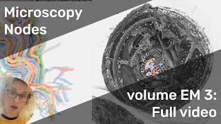
In this video, I will demonstrate how to load and show FIBSEM data using the Microscopy Nodes addon, now renamed from Tif2Blender. This tool allows for the generation of stunning 3D renders of microscopy images…
Use the Microscopy Nodes addon to generate stunning 3D renders of microscopy images. This video demonstrates how to transform volume electron microscopy data into a presentation-ready format, showcasing research on an…
In this video, I will show how to load segmentations in FIBSEM data using the Microscopy Nodes addon. This tool allows for generating beautiful 3D renders of microscopy images, enhancing visualization of environmental…
@MocaerK @SchwabYannick @PaoloRonchi10 and colleagues @EMBL: https://t.co/TU6H3ZmNKy
Dec 7, 2023 · 3 likes · 19,754 followersView post →
MK
Mocaer Karel
@MocaerK · X
RT @Co_Biologists: Thanks to Paolo Ronchi @PaoloRonchi10 for sharing his experience of APC-free #OA publishing in @J_Cell_Sci via our #Read…
Oct 18, 2023 · 6 shares · 112 followersView post →
AR
Alessandra Reversi
@AleReversi · X
RT @Co_Biologists: Thanks to Paolo Ronchi @PaoloRonchi10 for sharing his experience of APC-free #OA publishing in @J_Cell_Sci via our #Read…
Oct 18, 2023 · 6 shares · 318 followersView post →
ST
Shelly Turner
@shellyt2101 · X
RT @Co_Biologists: Thanks to Paolo Ronchi @PaoloRonchi10 for sharing his experience of APC-free #OA publishing in @J_Cell_Sci via our #Read…
Oct 18, 2023 · 6 shares · 131 followersView post →
PR
Paolo Ronchi
@PaoloRonchi10 · X
RT @Co_Biologists: Thanks to Paolo Ronchi @PaoloRonchi10 for sharing his experience of APC-free #OA publishing in @J_Cell_Sci via our #Read…
Oct 18, 2023 · 6 shares · 242 followersView post →
TB
The Company of Biologists
@Co_Biologists · X
Thanks to Paolo Ronchi @PaoloRonchi10 for sharing his experience of APC-free #OA publishing in @J_Cell_Sci via our #ReadAndPublish agreement with @embl @szilardlibrary
Read Paolo's paper https://t.co/dt6wGXgxzO
See if your institution also has an agreement https://t.co/WpJcxTt3x8 https://t.co/dV338C7OEn
Oct 18, 2023 · 10 likes · 6 shares · 15,432 followersView post →
RT @physorg_com: New #microscopy method can help put sharper focus on #plankton samples https://t.co/1txOyONnfn https://t.co/v6BAH5bx6I
Oct 6, 2023 · 13 shares · 1,788 followersView post →
JN
Jaroslav Novotný
@novotnyjarosl · X
RT @physorg_com: New #microscopy method can help put sharper focus on #plankton samples https://t.co/1txOyONnfn https://t.co/v6BAH5bx6I
Oct 6, 2023 · 13 shares · 717 followersView post →
P
Pisigomet
@pisigomet · X
RT @physorg_com: New #microscopy method can help put sharper focus on #plankton samples https://t.co/1txOyONnfn https://t.co/v6BAH5bx6I
Oct 6, 2023 · 13 shares · 289 followersView post →
DK
Dr. Larry B Keating
@DrLarryBKeating · X
RT @physorg_com: New #microscopy method can help put sharper focus on #plankton samples https://t.co/1txOyONnfn https://t.co/v6BAH5bx6I
Oct 6, 2023 · 13 shares · 818 followersView post →
AS
Ankit Singla
@In_AnkitSingla · X
RT @physorg_com: New #microscopy method can help put sharper focus on #plankton samples https://t.co/1txOyONnfn https://t.co/v6BAH5bx6I
Oct 6, 2023 · 13 shares · 1,480 followersView post →
JV
J C O V
@CesarVindell · X
RT @physorg_com: New #microscopy method can help put sharper focus on #plankton samples https://t.co/1txOyONnfn https://t.co/v6BAH5bx6I
Oct 5, 2023 · 13 shares · 730 followersView post →
MM
Manoj Mayogi Mishra
@mayogisense · X
RT @physorg_com: New #microscopy method can help put sharper focus on #plankton samples https://t.co/1txOyONnfn https://t.co/v6BAH5bx6I
Oct 5, 2023 · 13 shares · 574 followersView post →
S
sciencenews
@Scieducation1 · X
RT @physorg_com: New #microscopy method can help put sharper focus on #plankton samples https://t.co/1txOyONnfn https://t.co/v6BAH5bx6I
Oct 5, 2023 · 13 shares · 5,140 followersView post →
SS
Sagar Sinha
@Sagar_Sinha92 · X
RT @physorg_com: New #microscopy method can help put sharper focus on #plankton samples https://t.co/1txOyONnfn https://t.co/v6BAH5bx6I
Oct 5, 2023 · 13 shares · 503 followersView post →
CB
Christian Brey
@ChrisBrey1 · X
RT @physorg_com: New #microscopy method can help put sharper focus on #plankton samples https://t.co/1txOyONnfn https://t.co/v6BAH5bx6I
Oct 5, 2023 · 13 shares · 7,908 followersView post →
PO
Phys.org
@physorg_com · X
New #microscopy method can help put sharper focus on #plankton samples https://t.co/1txOyONnfn https://t.co/v6BAH5bx6I
Oct 5, 2023 · 32 likes · 13 shares · 1,022,459 followersView post →
JS
J Cell Science
@J_Cell_Sci · X
Issue 15 is complete!
Explore our ToC here: https://t.co/AWMjTYmode
On the cover: Segmentation of trichocysts in a three-dimensional rendering of the photosynthetic dinoflagellate Ensiculifera tyrrhenica; from Mocaer et al.:
https://t.co/N1W3zlecJ9 https://t.co/fU3Ig21Xko
Aug 16, 2023 · 15 likes · 19,754 followersView post →
VW
Venera Weinhardt
@VWein4 · X
RT @focalplane_jcs: This #EMmonday we are featuring the beautiful vCLEM paper from @MocaerK @SchwabYannick @PaoloRonchi10, et al. They reve…
Aug 15, 2023 · 7 shares · 343 followersView post →
DP
Daniel Yee PhD
@DanielPYee · X
RT @J_Cell_Sci: @MocaerK @SchwabYannick @PaoloRonchi10 @EMBL and colleagues reveal the 3D ultrastructure of a marine dinoflagellate using t…
Aug 14, 2023 · 10 shares · 131 followersView post →
K
KissedByElectrons
@ElectronKiss · X
RT @focalplane_jcs: This #EMmonday we are featuring the beautiful vCLEM paper from @MocaerK @SchwabYannick @PaoloRonchi10, et al. They reve…
Aug 14, 2023 · 7 shares · 420 followersView post →
KR
Kausthubh R
@kausthubh_r · X
RT @J_Cell_Sci: @MocaerK @SchwabYannick @PaoloRonchi10 @EMBL and colleagues reveal the 3D ultrastructure of a marine dinoflagellate using t…
Aug 14, 2023 · 10 shares · 664 followersView post →
SG
Seema Grewal
@seemasainson · X
RT @focalplane_jcs: This #EMmonday we are featuring the beautiful vCLEM paper from @MocaerK @SchwabYannick @PaoloRonchi10, et al. They reve…
Aug 14, 2023 · 7 shares · 1,658 followersView post →
MK
Mocaer Karel
@MocaerK · X
RT @J_Cell_Sci: @MocaerK @SchwabYannick @PaoloRonchi10 @EMBL and colleagues reveal the 3D ultrastructure of a marine dinoflagellate using t…
Aug 14, 2023 · 10 shares · 112 followersView post →
MK
Mocaer Karel
@MocaerK · X
RT @focalplane_jcs: This #EMmonday we are featuring the beautiful vCLEM paper from @MocaerK @SchwabYannick @PaoloRonchi10, et al. They reve…
Aug 14, 2023 · 7 shares · 112 followersView post →
PR
Paolo Ronchi
@PaoloRonchi10 · X
RT @focalplane_jcs: This #EMmonday we are featuring the beautiful vCLEM paper from @MocaerK @SchwabYannick @PaoloRonchi10, et al. They reve…
Aug 14, 2023 · 7 shares · 242 followersView post →
RH
Ricardo Henriques
@HenriquesLab · X
RT @focalplane_jcs: This #EMmonday we are featuring the beautiful vCLEM paper from @MocaerK @SchwabYannick @PaoloRonchi10, et al. They reve…
Aug 14, 2023 · 7 shares · 15,335 followersView post →
GD
Gautam Dey
@Dey_Gautam · X
RT @focalplane_jcs: This #EMmonday we are featuring the beautiful vCLEM paper from @MocaerK @SchwabYannick @PaoloRonchi10, et al. They reve…
Aug 14, 2023 · 7 shares · 8,184 followersView post →
F
FocalPlane
@focalplane_jcs · X
This #EMmonday we are featuring the beautiful vCLEM paper from @MocaerK @SchwabYannick @PaoloRonchi10, et al. They reveal the ultrastructure of a marine dinoflagellate, validating their method for targeting microorganisms from heterogenous field samples.
https://t.co/l3IAruUiBz https://t.co/tIxVwH3Beu
Aug 14, 2023 · 28 likes · 7 shares · 5,963 followersView post →
WZ
Weisong Zhao
@weisong_zhao · X
RT @J_Cell_Sci: @MocaerK @SchwabYannick @PaoloRonchi10 @EMBL and colleagues reveal the 3D ultrastructure of a marine dinoflagellate using t…
Aug 11, 2023 · 10 shares · 745 followersView post →
B
BioImagingUK
@BioImagingUK · X
RT @J_Cell_Sci: @MocaerK @SchwabYannick @PaoloRonchi10 @EMBL and colleagues reveal the 3D ultrastructure of a marine dinoflagellate using t…
Aug 11, 2023 · 10 shares · 4,573 followersView post →
رع
رمزي عمران
@Ramzi7004 · X
RT @J_Cell_Sci: @MocaerK @SchwabYannick @PaoloRonchi10 @EMBL and colleagues reveal the 3D ultrastructure of a marine dinoflagellate using t…
Aug 10, 2023 · 10 shares · 81 followersView post →
XE
X-Ray Imaging Team @EMBL
@HiTT_Hamburg · X
RT @J_Cell_Sci: @MocaerK @SchwabYannick @PaoloRonchi10 @EMBL and colleagues reveal the 3D ultrastructure of a marine dinoflagellate using t…
Aug 10, 2023 · 10 shares · 127 followersView post →
PR
Paolo Ronchi
@PaoloRonchi10 · X
RT @J_Cell_Sci: @MocaerK @SchwabYannick @PaoloRonchi10 @EMBL and colleagues reveal the 3D ultrastructure of a marine dinoflagellate using t…
Aug 10, 2023 · 10 shares · 242 followersView post →
OD
Omaya Dudin
@dudin_o · X
RT @J_Cell_Sci: @MocaerK @SchwabYannick @PaoloRonchi10 @EMBL and colleagues reveal the 3D ultrastructure of a marine dinoflagellate using t…
Aug 10, 2023 · 10 shares · 1,731 followersView post →
JS
J Cell Science
@J_Cell_Sci · X
@MocaerK @SchwabYannick @PaoloRonchi10 @EMBL and colleagues reveal the 3D ultrastructure of a marine dinoflagellate using their vCLEM workflow for targeting environmental microorganisms from heterogeneous samples.
Highlight: https://t.co/cRBKpjiMN2
T&R: https://t.co/N1W3zleKyH https://t.co/AjP22VNIv8
Aug 10, 2023 · 23 likes · 10 shares · 19,754 followersView post →
GM
Giulia Mizzon
@MizzonGiulia · X
RT @PaoloRonchi10: The final version of our manuscript on Targeted volume correlative light and electron microscopy of an environmental mar…
The final version of our manuscript on Targeted volume correlative light and electron microscopy of an environmental marine microorganism is out in JCS
@MocaerK @SchwabYannick @VolumeEM1 https://t.co/PUBKsSb7Ag
"We are planning to address the concern on technical variability in our revised version, together with a better explanation of time scales & automated VS manual aspect of the steps used for this workflow."
#ReviewedPreprints include the author's response
https://t.co/JwbB1XNdqz
'Targeted volume Correlative Light and Electron Microscopy of an environmental marine microorganism'
#ReviewedPreprint #ElectronMicroscopy #Microbiology
@MocaerK @PaoloRonchi10 et al @embl
https://t.co/JwbB1XNdqz https://t.co/eUweXvMnWn
May 31, 2023 · 6 likes · 5,258 followersView post →
RC
Review Commons
@ReviewCommons · X
RT @biorxivpreprint: #PeerReview from @ReviewCommons of
May 23, 2023 · 1 share · 5,258 followersView post →
 PR
 PO
 F
 JS
 TB


 MK
 AR
 ST
 DS
 NW
 JN
 P
 DK
 AS
 JV
 MM
 S
 SS
 CB
 VW
 DP
 K
 KR
 SG
 RH
 GD
 WZ
 B
 رع
 XE
 OD
 GM
 IF
 RR
 NS
 CB
 三投
 PB
 BG
 EH
 RC
 BB
 B
 BR