Synopsis of Social media discussions
The overall tone suggests strong recognition of the publication's importance, with some posts calling it 'the new Bible' and emphasizing its groundbreaking approach. Words like 'adapts itself' and mentions of advanced imaging techniques convey excitement about technological innovations. Additionally, references to related methods, such as machine learning applications, indicate active engagement with the content's broader implications in developmental biology.
Agreement
Moderate agreementMany references acknowledge the significance of the publication, with phrases like 'Here the new Bible' and the inclusion of the article's citation, indicating strong support or acknowledgment of its importance.
Interest
High level of interestSeveral discussions highlight curiosity about the methodology and implications, such as mentioning new tools like light-sheet microscopy and neural networks, showing active interest in the technological advancements.
Engagement
Moderate level of engagementSome posts go further by discussing related techniques like machine learning for cell analysis, sharing detailed observations, and referencing other researchers' work, suggesting a moderate level of deep engagement.
Impact
High level of impactThe enthusiasm expressed through hashtags like '#reproducibility', and references to the article as a 'new Bible' or indicating 'trend' scores, reflect a perception of high impact within the scientific or research community.
Social Mentions
YouTube
2 Videos
10 Posts
44 Posts
Blogs
8 Articles
News
18 Articles
2 Posts
Metrics
Video Views
839,319
Total Likes
11,309
Extended Reach
1,143,463
Social Features
84
Timeline: Posts about article
Top Social Media Posts
DNA&RNA Universe
In Toto Imaging and Reconstruction of Post-Implantation Mouse Development at the Single-Cell Level (2018)
https://t.co/ZMZfZroER8
source vid: https://t.co/1wE2YXI1mW
#science #biology #microscopy #fluorescence #FluorescenceFriday @_atanas_ @simongerman600 @grawoig @NovogeneLS https://t.co/NICzBeewiZ
view full post
Yara Sanchez Corrales
Twitter folks: do you know examples of using #ML #deeplearning to extract cell behaviour from images? I can only recall McDole et al., 2018 (https://t.co/oiuiBmCfHm), where a convolutional neural network (#CNN). was used to find diving cells.
Please RT. Thanks!
view full post
Dagmar Iber
We think that because of the low mesoderm area in caudal regions, the embryo requires #zippering to fully close its #neuraltube.
Movie with closure+zippering from our wonderful co-author Katie McDole, previously published in @CellCellPress: https://t.co/h3IivOsdWo https://t.co/z065Z5wxGt
view full post
Kay Schink
@ManuelTHERY @preibischs And this one from the Keller lab should have similar movies from mice :
https://t.co/LIfoxqlNeR
view full post
Adrián Candelas
Here the new Bible:
https://t.co/xoReaR5jPR
view full post
Posts referencing the article
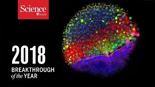
Breakthrough Scientific Achievements of 2018 Unveiled
Learn about our Breakthrough of the Year tracking development cell by cell. The study introduces a lightsheet microscope adapting to embryo changes, enabling detailed observation of early development stages like gastrulation and organ formation, along with advanced cell tracking tools.
December 20, 2018
824,922 views

In Toto Imaging of Post-Implantation Mouse Embryo Development
This timelapse video utilizes a specialized lightsheet microscope to observe mouse embryo development at the single-cell level, capturing cell behaviors during early stages like gastrulation and organ formation.
October 22, 2018
14,397 views
-

RT @dz_gong: Paper read today #275: In Toto Imaging and Reconstruction of Post-Implantation Mouse Development at the Single-Cell Level #Lig…
view full postFebruary 10, 2023
1
-
DZ Gong | 龚 | ゴン | 공
@dz_gong (Twitter)Paper read today #275: In Toto Imaging and Reconstruction of Post-Implantation Mouse Development at the Single-Cell Level #LightSheet 10.1016/j.cell.2018.09.031 A light-sheet microscope adapts itself to the changes in size, shape, and optical properties of the mouse embryo. https://t.co/xfcmrllz0E
view full postFebruary 9, 2023
1
1
-
fabrice senger
@fabrice_senger (Twitter)RT @adriancandelas: Here the new Bible: https://t.co/xoReaR5jPR
view full postOctober 18, 2022
1
-
Adrián Candelas
@adriancandelas (Twitter)One more… McDole et al., 2018 https://t.co/SkdwkLTSye
view full postOctober 18, 2022
-
Adrián Candelas
@adriancandelas (Twitter)Here the new Bible: https://t.co/xoReaR5jPR
view full postOctober 18, 2022
1
1
-
Jinglei Zhai, 晶磊 翟
@jingleidi (Twitter)RT @DagmarIber: We think that because of the low mesoderm area in caudal regions, the embryo requires #zippering to fully close its #neural…
view full postAugust 15, 2022
2
-
Susan Lowell de Solórzano
@1Biotensegrity (Twitter)RT @DagmarIber: We think that because of the low mesoderm area in caudal regions, the embryo requires #zippering to fully close its #neural…
view full postOctober 24, 2021
2
-
Dagmar Iber
@DagmarIber (Twitter)We think that because of the low mesoderm area in caudal regions, the embryo requires #zippering to fully close its #neuraltube. Movie with closure+zippering from our wonderful co-author Katie McDole, previously published in @CellCellPress: https://t.co/h3IivOsdWo https://t.co/z065Z5wxGt
view full postOctober 24, 2021
9
2
-
RRID Robot
@RobotRrid (Twitter)The authors of "In Toto Imaging and Reconstruction of Post-Implant…" (https://t.co/udYQMl9HVi) included RRIDs in their paper! Thank you for making your #methodsmatter. #reproducibility
view full postSeptember 6, 2021
-
Kay Schink
@kschink (Twitter)@ManuelTHERY @preibischs And this one from the Keller lab should have similar movies from mice : https://t.co/LIfoxqlNeR
view full postFebruary 12, 2021
2
-
Erika Tsingos
@tisrenk (Twitter)RT @DrYaraSanchez: Twitter folks: do you know examples of using #ML #deeplearning to extract cell behaviour from images? I can only recall…
view full postJanuary 28, 2020
9
-
Araújo Lab
@AraujolabUB (Twitter)RT @DrYaraSanchez: Twitter folks: do you know examples of using #ML #deeplearning to extract cell behaviour from images? I can only recall…
view full postJanuary 27, 2020
9
-
Will Hill
@DrWilliamHill (Twitter)RT @DrYaraSanchez: Twitter folks: do you know examples of using #ML #deeplearning to extract cell behaviour from images? I can only recall…
view full postJanuary 27, 2020
9
-
Assaf Zaritsky
@AssafZaritsky (Twitter)RT @DrYaraSanchez: Twitter folks: do you know examples of using #ML #deeplearning to extract cell behaviour from images? I can only recall…
view full postJanuary 27, 2020
9
-
A
@AnnaBajur (Twitter)RT @DrYaraSanchez: Twitter folks: do you know examples of using #ML #deeplearning to extract cell behaviour from images? I can only recall…
view full postJanuary 27, 2020
9
-
Luisma Escudero
@lmescu (Twitter)RT @DrYaraSanchez: Twitter folks: do you know examples of using #ML #deeplearning to extract cell behaviour from images? I can only recall…
view full postJanuary 27, 2020
9
-
Dr Matt Benton
@DrMattBenton (Twitter)RT @DrYaraSanchez: Twitter folks: do you know examples of using #ML #deeplearning to extract cell behaviour from images? I can only recall…
view full postJanuary 27, 2020
9
-
Yara Sanchez Corrales
@DrYaraSanchez (Twitter)Twitter folks: do you know examples of using #ML #deeplearning to extract cell behaviour from images? I can only recall McDole et al., 2018 (https://t.co/oiuiBmCfHm), where a convolutional neural network (#CNN). was used to find diving cells. Please RT. Thanks!
view full postJanuary 27, 2020
9
9
-
Sharer UsSharing
@SharerUssharing (Twitter)RT @DNA_RNA_Uni: In Toto Imaging and Reconstruction of Post-Implantation Mouse Development at the Single-Cell Level (2018) https://t.co/ZM…
view full postDecember 11, 2019
18
-
Dave Grawoig, Ph.D.
@grawoig (Twitter)RT @DNA_RNA_Uni: In Toto Imaging and Reconstruction of Post-Implantation Mouse Development at the Single-Cell Level (2018) https://t.co/ZM…
view full postDecember 11, 2019
18
-
Newyork Fitness
@NewYorkPass (Twitter)RT @DNA_RNA_Uni: In Toto Imaging and Reconstruction of Post-Implantation Mouse Development at the Single-Cell Level (2018) https://t.co/ZM…
view full postDecember 11, 2019
18
-
Dave Grawoig, Ph.D.
@grawoig (Twitter)RT @DNA_RNA_Uni: In Toto Imaging and Reconstruction of Post-Implantation Mouse Development at the Single-Cell Level (2018) https://t.co/ZM…
view full postNovember 18, 2019
18
-
Dave Grawoig, Ph.D.
@grawoig (Twitter)RT @DNA_RNA_Uni: In Toto Imaging and Reconstruction of Post-Implantation Mouse Development at the Single-Cell Level (2018) https://t.co/ZM…
view full postNovember 12, 2019
18
-
Lommond
@Lommond78 (Twitter)RT @DNA_RNA_Uni: In Toto Imaging and Reconstruction of Post-Implantation Mouse Development at the Single-Cell Level (2018) https://t.co/ZM…
view full postNovember 10, 2019
18
-
the reaper
@thparietallobe (Twitter)RT @DNA_RNA_Uni: In Toto Imaging and Reconstruction of Post-Implantation Mouse Development at the Single-Cell Level (2018) https://t.co/ZM…
view full postNovember 9, 2019
18
-
Scránny® Strange
@LAVOE_Jonnath (Twitter)RT @DNA_RNA_Uni: In Toto Imaging and Reconstruction of Post-Implantation Mouse Development at the Single-Cell Level (2018) https://t.co/ZM…
view full postNovember 9, 2019
18
-
DNA&RNA Universe
@DNA_RNA_Uni (Twitter)RT @DNA_RNA_Uni: In Toto Imaging and Reconstruction of Post-Implantation Mouse Development at the Single-Cell Level (2018) https://t.co/ZM…
view full postNovember 9, 2019
18
-
م. محمد العقيلي
@MohdAlaqeili (Twitter)RT @DNA_RNA_Uni: In Toto Imaging and Reconstruction of Post-Implantation Mouse Development at the Single-Cell Level (2018) https://t.co/ZM…
view full postNovember 9, 2019
18
-
Alice Lee
@alee7095 (Twitter)RT @DNA_RNA_Uni: In Toto Imaging and Reconstruction of Post-Implantation Mouse Development at the Single-Cell Level (2018) https://t.co/ZM…
view full postNovember 9, 2019
18
-
International Natural Product Sciences Taskforce
@_INPST (Twitter)RT @DNA_RNA_Uni: In Toto Imaging and Reconstruction of Post-Implantation Mouse Development at the Single-Cell Level (2018) https://t.co/ZM…
view full postNovember 9, 2019
18
-
Atanas G. Atanasov
@_atanas_ (Twitter)RT @DNA_RNA_Uni: In Toto Imaging and Reconstruction of Post-Implantation Mouse Development at the Single-Cell Level (2018) https://t.co/ZM…
view full postNovember 8, 2019
18
-
Dave Grawoig, Ph.D.
@grawoig (Twitter)RT @DNA_RNA_Uni: In Toto Imaging and Reconstruction of Post-Implantation Mouse Development at the Single-Cell Level (2018) https://t.co/ZM…
view full postNovember 8, 2019
18
-
John Paul Tosoc, PhD
@meJPAUL (Twitter)RT @DNA_RNA_Uni: In Toto Imaging and Reconstruction of Post-Implantation Mouse Development at the Single-Cell Level (2018) https://t.co/ZM…
view full postNovember 8, 2019
18
-
DNA&RNA Universe
@DNA_RNA_Uni (Twitter)In Toto Imaging and Reconstruction of Post-Implantation Mouse Development at the Single-Cell Level (2018) https://t.co/ZMZfZroER8 source vid: https://t.co/1wE2YXI1mW #science #biology #microscopy #fluorescence #FluorescenceFriday @_atanas_ @simongerman600 @grawoig @NovogeneLS https://t.co/NICzBeewiZ
view full postNovember 8, 2019
27
18
-
yy26
@yy26 (Twitter)In toto imaging and reconstruction of post-implantation mouse development at the single-cell level https://t.co/36KWRNlmw0
view full postMarch 5, 2019
-
sayum
@szayu1 (Twitter)In Toto Imaging and Reconstruction of Post-Implantation Mouse Development at the Single-Cell Level https://t.co/Va2TKgC27v
view full postJanuary 12, 2019
-
the paper link
@the_paper_link (Twitter)[trend] up+11: *McDole K* et al (Cell) **In Toto Imaging and Reconstruction of Post-Implantation Mouse Development at the Single-Cell Level.** https://t.co/y0p4XjRzNt
view full postJanuary 9, 2019
-
the paper link
@the_paper_link (Twitter)[trend] up+5: *McDole K* et al (Cell) **In Toto Imaging and Reconstruction of Post-Implantation Mouse Development at the Single-Cell Level.** https://t.co/y0p4XjRzNt
view full postJanuary 8, 2019
-
the paper link
@the_paper_link (Twitter)[trend] up+9: *McDole K* et al (Cell) **In Toto Imaging and Reconstruction of Post-Implantation Mouse Development at the Single-Cell Level.** https://t.co/y0p4XjRzNt
view full postJanuary 6, 2019
-
the paper link
@the_paper_link (Twitter)[trend] up+5: *McDole K* et al (Cell) **In Toto Imaging and Reconstruction of Post-Implantation Mouse Development at the Single-Cell Level.** https://t.co/y0p4XjRzNt
view full postJanuary 5, 2019
-
the paper link
@the_paper_link (Twitter)[trend] up+3: *McDole K* et al (Cell) **In Toto Imaging and Reconstruction of Post-Implantation Mouse Development at the Single-Cell Level.** https://t.co/y0p4XjRzNt
view full postJanuary 4, 2019
-
the paper link
@the_paper_link (Twitter)[trend] up+4: *McDole K* et al (Cell) **In Toto Imaging and Reconstruction of Post-Implantation Mouse Development at the Single-Cell Level.** https://t.co/y0p4XjRzNt
view full postJanuary 3, 2019
-
the paper link
@the_paper_link (Twitter)[trend] up+20: *McDole K* et al (Cell) **In Toto Imaging and Reconstruction of Post-Implantation Mouse Development at the Single-Cell Level.** https://t.co/y0p4XjRzNt
view full postJanuary 2, 2019
-
the paper link
@the_paper_link (Twitter)[trend] up+14: *McDole K* et al (Cell) **In Toto Imaging and Reconstruction of Post-Implantation Mouse Development at the Single-Cell Level.** https://t.co/y0p4XjRzNt
view full postJanuary 1, 2019
Abstract Synopsis
- This study introduces a special lightsheet microscope that can adapt to changing mouse embryo sizes and shapes, allowing scientists to watch cell behavior during early development stages like gastrulation and organ formation.
- The researchers also created advanced computer tools to track cell movements, divisions, and tissue changes over time within the embryo, building a detailed dynamic map of development.
- By analyzing multiple embryos together, they made a comprehensive "dynamic atlas" of postimplantation mouse development, which serves as a valuable resource for understanding how tissues and organs form.
Petra Kučerová
@Peta_Kucerova (Twitter)